Explain the Differences Between the Layers of the Gastrointestinal Tract
Explain how the structure of each of the following digestive tract compartme. The structure of these layers varies in different regions of the digestive system depending on their function.
Printed From Student Consult Berne And Levy Physiology 6e The Online Medical Library For Students Plus Usmle Steps 123 Ver 2 9
Start studying Diet Digestion.

. An anatomical model of the human stomach is an example of the models at organ level while at the tissue level the different muscle layers and their cell types. Oesophagus is exceptional in having a compound squamous epithelium on the inside and an adventitia on the outside. In certain regions the mucosa develops folds that increase the.
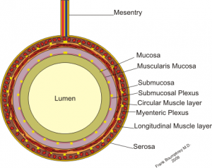
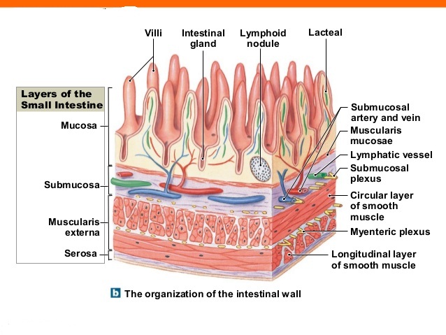
Illustration of the different spatial scales of models involved in the representation of the gastrointestinal system. The GI tract contains four layers. The muscularis mucosa is a thin layer of smooth muscle that supports the mucosa and.
These layers are named as. Biology questions and answers. The inner layer is circular and the outer layer is longitudinal.
An anatomical torso geometry is represented at the broadest scale. It lines the lumen of the digestive tract. The organs of the gastrointestinal tract contain layers of muscles.
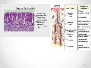
The wall of the stomach consists of the usual four layers present in other parts of the gastrointestinal tract. The mucosa is relatively thick and contains numerous tubular glands. All segments of the GI tract are divided into four layers.
The outer layer of the stomach wall is smooth continuous with the parietal peritoneum. These include most of the stomach first part of the duodenum all of the small intestine caecum and appendix transverse colon sigmoid colon and rectum. In these sections of the gut there is a clear boundary between the.
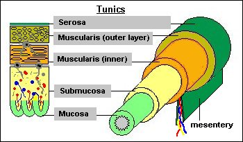
The four layers in more detail. There are 4 layers in gastrointestinal tract. This is the simplified version.
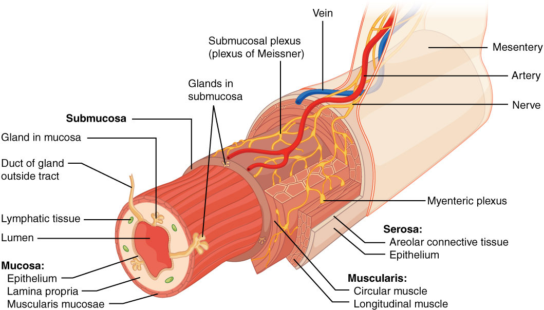
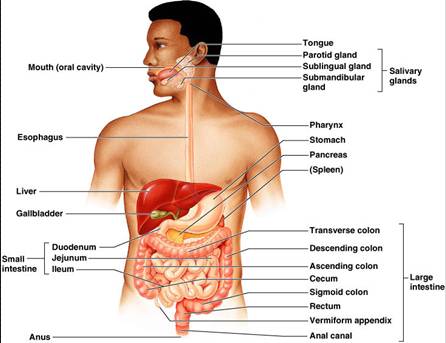
In mammalian alimentary canal we can see four layers almost in all organs in gastrointestinal tract with minor and major differences in different organs. Between the two muscle layers are the myenteric or Auerbachs plexus. The digestive system is a broader term that includes other structures including the accessory organs of digestion such as the liver gallbladder and pancreas.
There are usually two layers. We review their content and use your feedback to keep the quality high. Composed of 3 layers epithelial areolar connective smooth muscle Contains lymph tissue.
Explain the differences between the layers of the gastrointestinal tract. The muscularis externa of the large intestine is different from that of the small intestine in that the outer longitudinal layer of smooth muscle varies in thickness and forms three thick longitudinal bands the taeniae coli taenia worm. Four layers of the Gastointestinal Tract.
The submucosa is relatively thick highly vascular and serves the. These layers of smooth muscle are used for peristalsis rhythmic waves of contraction to move food down through the gut. Lining of the lumen.
The fact is that there are more sublayers. 05 points Answer. Serous layer or serosa.
The outermost layer of the gastrointestinal tract consists of several layers of connective tissue. Human Anatomy Lab Manual. Serosa Outermost layer 2.
The muscularis mucosae is also thick and in some areas it consists of 3 layers of smooth muscle although this layering is not always visible. The gastrointestinal tract is made up of a series of hollow organs joined together in a long tube with many folds from the mouth to the anus. Contains the submucosal enteric neural plexus that controls GI secretions and localized blood flow.
The innermost layer is the mucosa underneath this is the submucosa followed by the muscularis propria and finally the outermost layer - the adventitia. Intraperitoneal parts of the GI tract are covered with serosa. A bolus of food enters the stomach from the esophagus.
Muscularis propria made up of muscles 3. The GI tract is composed of four layers. The Layers of the GI tract.
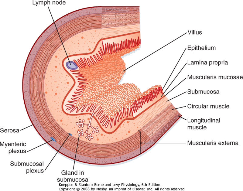
The hollow organs that make up the gastrointestinal tract include the mouth oesophagus stomach small intestine cecum colon large intestine rectum and anal canal. Learn vocabulary terms and more with flashcards games and other study tools. The gastrointestinal GI tract is formed with a few exceptions by four concentric layers of tissue.
Summarize the role of GI tract hormones following a meal. These are from deep to superficial the mucosa submucosa muscular or muscularis and the serosa layers. From inside to outside these are mucosa submucosa muscular layer and serosa.
The stomach has an extra layer an inner oblique muscular layer. The mucosa surrounds the lumen of the GI tract and consists of an epithelial cell layer supported by a thin layer of connective tissue known as the lamina propria. The mucosa or mucous membrane layer is the innermost tunic of the wall.
Explain the differences between the layers of the gastrointestinal tract. The inner wall mucosa and submucosa layers is thrown into folds known as rugae or gastric folds which allow the stomach to distend upon the entry of the food. The muscle of the inner layer is arranged in circular rings around the tract whereas the muscle of the outer layer is arranged longitudinally.
The mucosa consists of epithelium an underlying loose connective tissue layer called lamina propria and a thin layer of smooth muscle called the muscularis mucosa. Adventia layer or serosa outermost layer of loose connective tissue - covered by the visceral peritoneum. Gastrointestinal tract is an organ system in humans and other animals that take in food digest it absorb nutrients and expel it out in the form of feces Gastrointestinal Tract Diagram The gastrointestinal diagram given below represents the different parts of the tract that include the oral cavity oesophagus stomach intestines and.
33 Explain how differences in the structure of the gut wall on the oesophagus stomach and ileum are related to their roles in digestion. The mucosa layer is characterized by the presence of. Contains many glands which open into the lumen by way of ducts.
The innermost layer is mucosa epithelium lamina propria and muscular mucosaeouter to mucosa is the submucosa then muscularis propria inner circular muscle layer intermuscular space a. This tract consists of the stomach and intestine and sometimes includes all the structures from the mouth to the anus. This section happened to be cut such that a piece of one of these longitudinal bands may be seen.
Each layer has different tissues and functions. From the inside out they are called. Explain the differences between the layers of the gastrointestinal tract.

Layers Of The Alimentary Canal Boundless Anatomy And Physiology

Layers Of The Gi Tract Adapted From 23 Download Scientific Diagram
From The Lumen Outward What Are The Layers Of The Gastrointestinal Tract Socratic
Digestive System Histology
Microanatomy Of The Digestive Tube

Difference Between Muscularis Layer Of Esophagus And Stomach Compare The Difference Between Similar Terms
Histology Of Gastrointestinal Tract
From The Lumen Outward What Are The Layers Of The Gastrointestinal Tract Socratic

Anatomy And Physiology Of The Gastrointestinal System Notes Diagrams Illustrations Osmosis

Gi Tract
Is There A Difference Between The Digestive Tract And The Gastrointestinal Tract Socratic
What Are The Layers Of The Gastrointestinal Tract What Are Their Functions Socratic

Gastrointestinal Tract 1 The Mouth And Oesophagus Nursing Times

Layers Of The Alimentary Canal Boundless Anatomy And Physiology

Relationship Between Histological And Ultrasonographic Gastrointestinal Download Scientific Diagram
2

Oral The Histology Guide

Difference Between Muscularis Layer Of Esophagus And Stomach Compare The Difference Between Similar Terms

Illustrated Gastric Stomach Layers 3 Layers Of The Stomach Anatomy


Comments
Post a Comment